Tarsal Coalition is defined as a congenital union or failure of segmentation between 2 or more tarsal bones. The coalition may be osseous, cartilaginous or fibrous. It may occur at sites of normal articulation or may connect joints that do not usually articulate.The incidence is thought to be less than 1% with 50% being bilateral. Male patients are affected twice as often. The commonest site is the calcaneo-navicular joint (60%) with the middle facet of the talo-calcneal joint being next most common (30%).
Symptoms develop in the 2nd decade when ossification ofthe pre-existing fibrous or cartilaginous bar occurs. The calcaneo-navicular bar ossifies earlier and therefore becomes symptomatic around age 8 – 12. Talo-calcaneal symptoms develop around 12 – 16 years of age. Pain is activity related and may be felt around the sinus tarsi, anterolateral calf or over the sustentaculum tali. There may be a history of repeated ankle sprains. The forefoot is abducted and the hindfoot is usually in valgus. Peroneal muscle spasm is sometimes present. Subtalar motion is reduced or absent.
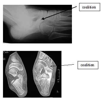
Plain x-rays may not demonstrate cartilaginous or fibrous bars. Talo-calcaneal coalition is best seen on the Harris 45 degree axial view. Indirect clues include talar beaking, subtalar joint narrowing -esp post facet, altered shape to the talar head and broadening of the lateral talar process. Calcaneo-navicular coalition is best seen with a 45 degree oblique view. An abnormal elongation to the anterior process of the calcaneus is present (ant eater nose sign). There may also be hypoplasia of the talar head and flattening of the navicular as it approaches the calcaneus.
The talo-calcaneal joint is best seen on coronal CT. Measurement of coalition size is not standardised. It can be estimated as a percentage of anterior/middle facet or posterior facet or total joint area. Hindfoot valgus should also be measured. Calcaneo-navicular coalition is best demonstrated with transverse CT slices. MRI scanning correlates well with CT findings and fibrous coalitions are well seen. Bone scanning may help to diagnose exclude other conditions such as infection, occult fracture, inflammatory arthritis or tumour.
Asymptomatic cases do not require treatment. Patients with mild pain can be managed non-operatively with analgesia, activity modification, medial heel wedge or plastizoate shoe insert. For more persistent symptoms, 4 weeks in a walking short leg cast may be beneficial. Surgery should be considered for moderate and severe symptoms that have not responded to conservative therapy.
Calcaneo-navicular coalitions are best managed by bar resection and interposition with fat or muscle. The foot is immobilised in a plaster cast for 3 weeks postoperatively. The overall results are good – (90%).
Treatment for talocalcaneal coalition is more controversial. Previously arthrodesis was considered the best option if conservative treatment failed. The recent literature is reporting good results with resection of the coalition and this is now considered the procedure of choice. Good to excellent results are seen if the coalition occupies <1/3 total subtalar joint area or <1/2 area of Post facet area. Heel valgus < 16 – 21 degrees has a better prognosis. Calcaneal osteotomy helps to recentre the heel, realign the weight bearing axis and relieve ligamentous strain. Triple arthrodesis is offered as a salvage option in the skeletally mature foot.
Treatment for talocalcaneal coalition is more controversial. Previously arthrodesis was considered the best option if conservative treatment failed. The recent literature is reporting good results with resection of the coalition and this is now considered the procedure of choice. Good to excellent results are seen if the coalition occupies <1/3 total subtalar joint area or <1/2 area of Post facet area. Heel valgus < 16 – 21 degrees has a better prognosis. Calcaneal osteotomy helps to recentre the heel, realign the weight bearing axis and relieve ligamentous strain. Triple arthrodesis is offered as a salvage option in the skeletally mature foot.
 Dr. Leonard kuo