
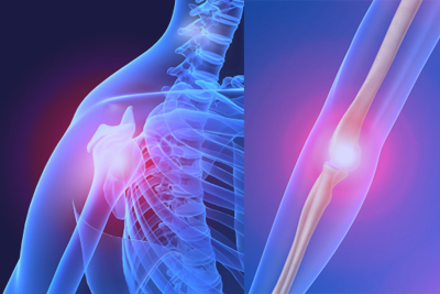
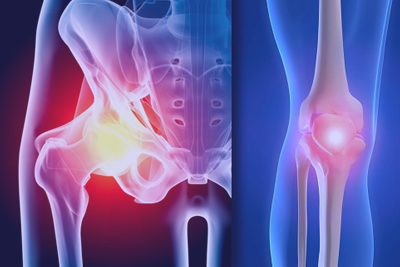

In this condition a segment of cartilage with its underlying subchondral bone separates from the articular surface of synovial joints. The incidence is about 1 : 5000 in the knee with 30% being bilateral. Males are affected three times more commonly than females. There are two groups affected – children / adolescents and young adults. In the knee, the medial femoral condyle is involved in 80-85% of cases and the lesion may be located inferolaterally, central or centro-lateral. The lateral condyle may be affected inferocentral or posterior. OCD of the talus affects the posteromedial -55% or anterolateral corner - 45%.
The cause is unknown but is probably multifactorial. Theories include trauma, ischaemia, familial factors, secondary to accessory ossification and endocrinopathy. Primary pathological changes occur in bone with secondary changes developing in the cartilage. There is focal ischaemia and subsequent avascular necrosis with the development of an osteochondral fragment. Variable attempts at repair occur with revascularisation and creeping substitution leading to healing. If this response is poor, a fibrous nonunion develops and the fragment may separate. The overlying cartilage is nourished by synovial fluid and is initially normal and continues to grow. Later there is loss of support, softening and eventual degeneration. Lesions may therefore be attached, detached or loose.
Patients may present with a history of vague joint discomfort and intermittent swelling. Symptoms tend to be related to activity and exercise. Later clicking, giving way and locking from a loose body may occur. The diagnosis is often missed in the ankle initially. It should be suspected if there is a history of lateral ankle sprain and persistent symptoms such as pain and swelling.
Examination may reveal wasting, an antalgic limp and local tenderness. Look for an effusion and loss in joint motion. Wilsons sign may be positive in the knee.
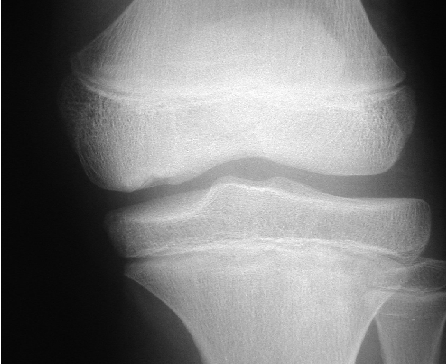
Xrays of the knee should include an intercondylar notch view. A crescent shaped area of radiolucency in subchondral bone may be visible. In the ankle, 1/3 of lesions are missed. Mortise views in varying degrees of PF and DF may assist. CT scanning is useful in assessing the location, size, stage and healing of the lesion. Arthrography has been replaced by MRI which is excellent for assessing the state of the overlying cartilage. Bone scan is helpful if the diagnosis is unclear and helps to distinguish from anomalous ossification centres.
Xrays of the knee should include an intercondylar notch view. A crescent shaped area of radiolucency in subchondral bone may be visible. In the ankle, 1/3 of lesions are missed. Mortise views in varying degrees of PF and DF may assist. CT scanning is useful in assessing the location, size, stage and healing of the lesion. Arthrography has been replaced by MRI which is excellent for assessing the state of the overlying cartilage. Bone scan is helpful if the diagnosis is unclear and helps to distinguish from anomalous ossification centres.
The aim of treatment is to prevent detachment of the fragment, improve blood supply and promote healing. Considerations in management include age – patients with open physes do better. Under the age of 12, lesions often heal spontaneously but this may take up to 2 years. A variable outcome is seen over the age of 12. Other considerations are anatomical site -weight bearing vrs non weight bearing location, size of lesion and stage.
Non operative management for symptomatic lesions in the knee involves activity restriction, a period of immobilisation and quadriceps rehabilitation. X-rays are taken 3-4 monthly to monitor progress.
Arthroscopy allows direct visualisation of the lesion and is useful for both diagnostic and therapeutic purposes. Symptomatic but undetached lesions that are not healing can be drilled with smooth K wires to encourage revascularisation. Detaching lesions are managed by debridement, drilling and in situ pinning with divergent K wires, headless compression screws or bio absorbable pins. If possible, fully detached lesions should also be internally fixed. Unsalvageable loose bodies are removed and the crated debrided and drilled. Salvage options include mosaicplasty, osteochondral allografting and osteotomy.
OCD involving the ankle is managed along similar lines. Asymptomatic Berndt and Harty stage 1,2 and 3 lesions require no treatment. Symptomatic stage 1 and 2 lesions are initially managed nonoperatively. Failing this, arthroscopic surgery is indicated. Intact lesions are treated by multiple drilling preferably from a non weight bearing surface. Partially detached and undisplaced detached areas require debridement, drilling and fragment fixation. Detached and loose lesions need ORIF if large and acute. Small and unsalvageable fragments are excised.
The prognosis is best if the defect is small (<1cm), involves a NWB surface, is in an earlier stage and the lesion heals before physeal closure.
 Dr. Leonard kuo