Introduction
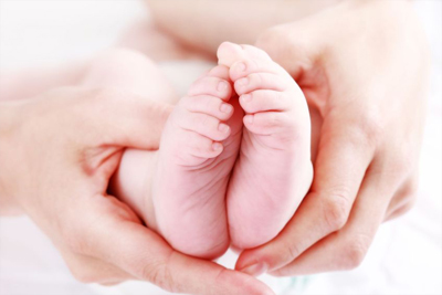
Leg length discrepancy is a fairly common problem that affect children of differing ages. It is seen in a wide variety of conditions and associated with certain problems.
Leg length discrepancy may contribute to
1) Increased energy expenditure in gait.
2) Cosmetically disturbing gait
3) Need for a prosthesis or shoe lift
4) Contracture of ankle
5) Scoliosis (Curvature of the spine)
6) Low back problems
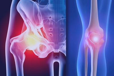
7) Late degenerative OA hip
Causes:
There are 3 categories of leg length discrepancy.
a) Those causing difference in length but not in growth rate - eg Fracture with overlap
b) Those that are equal in length initially but difference in growth rate leads to leg length discrepancy - eg radiation, physeal arrest
c) Those that do both - eg congenital conditions/ various reduction defects
Thus it is important to consider not only the initial leg length discrepancy at start of treatment but also the differential in growth rates. In congenital conditions, the growth inhibition is a fixed percentage of normal side. This enables the surgeon to give a good estimate of the ultimate leg length difference.
List of causes of leg length discrepancy.
| Shortening | Lengthening |
Congenital: Hemiatrophy Skeletal Dysplasia |
Hemihypertrophy Vascular Malformation |
| Infection: Growth Plate damage |
Metaphyseal Osteomyelitis Septic Arthritis |
| Paralysis: Poliomyelitis |
|
| Tumours: Enchondromatosis Osteochondromatosis |
Haemangioma Neurofibromatosis |
| Trauma: Physeal damage Overriding fractures |
Healing fractures Diaphyseal operations |
| Other: Perthes Disease SCFE Radiation therapy |
Clinical Assessment
Assessment of the whole patient is vital. It helps to detect factors that may have a bearing on your treatment goals. For example, factors such as neurological deficit, pelvic obliquity and fixed scoliosis may mean that perfect equalisation of leg lengths may not be ideal. Patients with weakness are better off having leg slightly shorter to allow ground clearance during swing phase eg foot drop. In fixed scoliosis, aim for a vertical lumbar spine rather than level pelvis.
Initially examination is carried out with the patient standing. Assess the level of knees, iliac crests, spine and size of the feet. Use blocks under the short leg to produce the optimal position of the legs and trunk. This shows the functional leg length discrepancy which in the case of fixed pelvic obliquity or spinal deformity may be different from the actual measured leg length discrepancy. The patient should be asked to walk and their gait observed. Look for weakness eg hemiplegic gait, trendelenburg or foot drop.
With the patient lying, measure apparent leg length discrepancy (umbilicus to MM) and real leg length discrepancy (ASIS to MM) or to foot if shortening is below the ankle. Exclude fixed deformity in lower limbs eg hip / knee and assess joint stability.
Radiological assessment
This provides the most important information for decision making. Proper follow up of a child with leg length discrepancy requires regular xrays to monitor and obtain a comprehensive radiological record of past growth. The pattern of growth is most accurately defined by the relationships between leg length and skeletal age. This is more reliable than historical information such as heights of siblings or parents. Data on both variables is collected as measuring the leg length alone is not useful in predicting future growth. Separate Xrays are taken for determining 1) leg length and skeletal age (bone age). This is performed yearly early on, then more frequently as time for corrective surgery approaches.
a) Bone age is determined by comparing an xray of the left hand with standards in the Greulich and Pyle Atlasb) Leg lengths are determined by:3 foot standing films or CT Scannogram.
c) In complex deformities- Standing AP pelvis / hips and lumbar spine with leg length corrected with blocks allows a determination of functional discrepancy to aid in setting treatment goals.
Management
Decision making in leg length discrepancy requires an assessment of
a) Patient expectations - some find a shoe lift unacceptable
b) Determining the current leg length discrepancy
c) Estimating height of patient at maturity- shorten or lengthen leg
d) Predicting future growth as treatment will be determined largely by this. The most important information required is the anticipated leg length discrepancy at maturity and not the current discrepancy.
Prediction of future growth
Most conditions show constant growth inhibition during the growth years but some have variable effects on growth. Therefore it may not be possible to be entirely accurate in predicting ultimate leg length discrepancy. Eg. Juvenile Rh A / Certain endocrine and metabolic disorders. Other considerations include asymmetric slowing leading to angular deformities that may also need to be addressed.
Three methods are commonly used to monitor and predict future growth: It may be calculated mathematically using 1)Menelaus Method
2)Anderson Method
or determined graphically using the
3)Moseley Straight line graph
Menelaus Method
This is an empirical method and simple to apply. It uses chronological age to determine how many years are left for growth but relies on chronological age to be within 18 months of bone age. He makes certain assumptions.
1)Physeal fusion occurs at chronological age 16 in boys and age 14 in girls
2) Growth at physes occurs at a constant rate
3) Lower femur provides 3/8 inch (10mm) growth per year
4) Proximal tibia provides 2/8 inch (6mm) growth per year
5) Proximal femur and distal tibia provide 1/8 inch (3mm) growth per year
Anderson Method
: Plots skeletal age against leg length
:Uses further calculations and data from Anderson / Green / Messner growth remaining charts to ascertain
-future growth of long leg / short leg
-future increase in discrepancy
-ultimate discrepancy at maturity
-effect of epiphyseodesis and skeletal age and when this should be done
Moseley Method
Data may be recorded in tabular form, but this makes it difficult to understand growth patterns. It takes time and requires repeated analysis at each visit and may lead to mathematical errors. Data is easier to interpret if it is in graphical form. The Moseley Straight line graph method provides such a means of doing this. It uses the same data as the Green-Anderson method but converts the leg length mathematically to a linear relationship. By plotting leg length and skeletal age on different axes we gain an immediate representation of
: Growth percentile and skeletal maturity of pt.
: Past growth: allows us to predict future growth and ultimate leg length discrepancy
: enables assessment of the effects of corrective surgery such as epiphyseodesis and lengthening by altering the slope or position of the growth lines.
: avoids mathematical errors.
Treatment
This is dependent on the estimated leg length discrepancy at maturity. If it is anticipated to be:
1) <2cm: No treatment is required. It is generally well tolerated and no long term problems or disabilities result.
2)>2cm: operative or non-operative treatment
-treatment is considered if the child is unable to compensate adequately for the discrepancy eg. fixed pelvic tilt
-The child must be aware that there may still be a limp and the limb will not look entirely normal.
Non-operative Management
All leg length discrepancy greater than 2cm may be managed non-operatively although this obviously may not be the preferred option
1) 2-5cm: heel lift - should be 1cm less than discrepancy and can be tapered to a lesser lift at the sole
2) >5cm: heel lifts >5cm poorly tolerated as ankle stability compromised and ligament sprains occur.
: use Patten and boot or extension prosthesis - moulded polypropylene splint
Operative Management
1) If growth inhibition is due to partial but not complete damage to the growth plate, eg infection / trauma, it is sometimes possible to restore the growth rate of the leg with an epiphyseolysis.
2) Shorten long leg
-This is preferred if the leg length discrepancy is <5-6cm
a)Acute: Shortening Osteotomy
b)Gradual: Epiphyseodesis (Modulation of growth)
3) Lengthen short leg
-for leg length discrepancy > 6cm
-It would seem logical to adopt this method as it aims to make the abnormal leg more normal (though never completely normal) and leaves the normal leg alone.
BUT: the complications and morbidity associated with leg lengthening is considerable. It requires prolonged hospitalisation, multiple procedures, and multiple visits for follow up. Up to 20% of the starting length of the bone can be achieved. Any more is much more difficult and increases the complication rate. Multiple lengthenings may also be required.
4) Amputation
This may be considered if there is gross shortening, instability, a poor foot and leg lengths cannot be equalised. A prosthesis gives better function and cosmesis. The results are predictable and follow up requirements are much reduced.
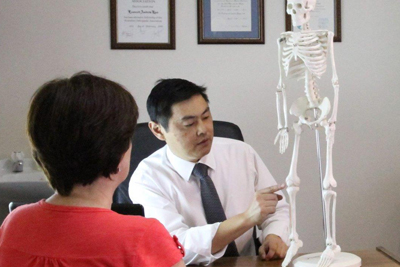
 Dr. Leonard kuo