

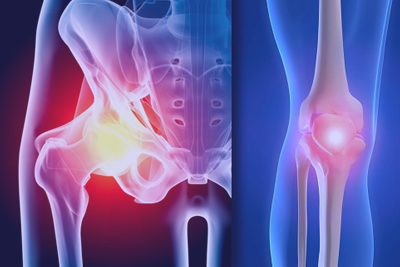
 Angular deformities of the Lower Limb in Children
In the assessment of children with angular deformities, the most important question to answer is “what is the natural history?” This may be obvious and based on an understanding of the underlying condition or it may not be known immediately and regular follow up will be required before this information becomes available. Broadly, there are two categories of angular deformities, those that are static and do not change and those that are dynamic which may either improve or progress with time. Examples of static deformities include malunited fractures at or near the end of growth or after surgery. These deformities may be treated anytime but there is usually no reason to defer treatment.
Deformities that tend to improve include physiological conditions such as genu varum, and genu valgum. Tibial valgus after proximal metaphyseal fracture also tends to improve and should be allowed the chance to do so. Surgical correction is considered only when no further improvement is seen and the deformity remains unacceptable.
Deformities that tend to improve include physiological conditions such as genu varum, and genu valgum. Tibial valgus after proximal metaphyseal fracture also tends to improve and should be allowed the chance to do so. Surgical correction is considered only when no further improvement is seen and the deformity remains unacceptable.
Progressive deformities require a less aggressive approach. Children can withstand significant angular deformities without sustaining joint damage or developing pain. In general the deformity should be tolerated for as long as possible to minimise the number of surgical corrections and hospitalisations. However there are exceptions which will be discussed. Pathological indicators include asymmetry, short stature, positive family history, deformity not following the physiological natural history and associated ligamentous laxity. Progressive disorders include blounts disease, physeal trauma, infection, metabolic and endocrine conditions and bone dysplasias and tumorous conditions. Congenital angular deformities such as tibial bowing have a variable natural history which is dependent upon the direction of the bow apex. Anterolateral bowing poses the greatest challenge with the tendency to fracture and subsequent development of pseudarthrosis.
Clinical assessment should include measurement of height and weight, assessment of rotational profile, joint stability and site of deformity in addition to the recording of the tibial-femoral angle and intermalleolar or intercondylar distances.
There are no long term prospective studies that have followed children with angular deformities to determine whether osteoarthritis is related to the magnitude of the angular deformity though several studies on angulation after malunited fractures have suggested an association with premature degenerative arthritis. The decision to correct deformity is not based on hard scientific evidence and based more on consideration of cosmesis or an intuitive feeling that the magnitude of the deformity will cause problems later. As a guide, angular deformities greater than 15 degrees should be considered for treatment.
The aims of treatment are to correct at or near the site of deformity, restore the mechanical axis and maintain a horizontal joint line. Options include hemi-epiphyseodesis using staples, screws or plates, osteotomy and gradual correction using external fixation.
 Dr. Leonard kuo